


|
|
|
|
|
|
|
|
|
||
|
EXPERIMENTAL PLAN |
|||
|
Principal Investigator/Program Director Williams, Robert W. |
|||
|
3. Changes to the Interface
To better understand some of the changes necessary to the interface,
it will perhaps be useful to discuss the current interface of the MBL
and then point out how we plan to change it. Screen shots will be
included, but the reader is encouraged to log into <nervenet.org/mbl/mbl.html>
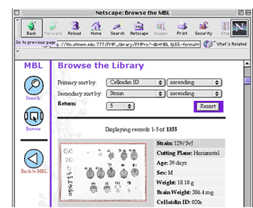
to view the current interface in detail. Upon entering the MBL home page (screen shot, right), one can choose to browse the collection of images from the brains in the collection, linked to the Brain Atlas <nervenet.org/mbl/mbl_main/atlas170.html>, the brain databases <nervenet.org/mbl/mbl_main/mbl_databases.html>, a table of strains <nervenet.org/mbl/mbl_main/mbl_straintable.html>, the iScope <http://nervenet.org/mbl/iscope/aboutiscope.html>, or other links in the library <nervenet.org/mbl/mbl_main/mbl_links.html>. Because most of the redesign of the interface of the MBL will involve the ways to view the photographic images taken from sections in the MBL, we will concentrate here on exploring this aspect of the current interface. Clicking on "Browse the Library" takes the user to the
interface for the FileMaker Pro image database (screen shot, right).
There, users can browse or search on a variety of criteria, including
strain, ID#, sex, age, brain weight, and body weight. All the cases
matching the search criteria appear in the rightmost frame. Clicking on
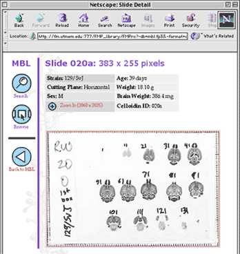
one of these thumbnails takes the user to a larger thumbnail of case
(383 255 pixels) with its strain, cutting
plane, sex, age, weight, brain weight, and ID# listed (screen shot
below, left).
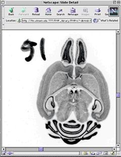
Clicking on this thumbnail then takes the user to the full 3060
2036 pixel image with a resolution of 25 µm/pixel. An example of one
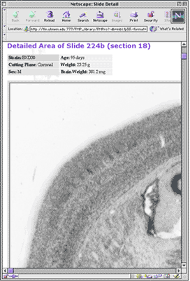
section of this image is shown in the screen shot on the right. C Improve Navigation from Slide to Images Our current methods for moving from the thumbnail of a slide to an image of an individual section are rather clumsy. We propose to implement the ability to click on any section in the thumbnail to immediately call up the higher-resolution image of that section. One way to think of this would be as if we were using the thumbnail as an image map on a web page. While the idea behind this change in the interface is the same, our methods of implementation will be different, as we will have sophisticated links in place between all the images in a given brain. Create JavaScript and Java Applets Tools and Techniques that Allow for Faster and More Transparent Navigation Through the MBL Collection. The current methods of navigation through the MBL are serviceable but not yet optimized. During the course of the proposal, we will create a variety of tools that will expedite the most common requests of users of the MBL. For example, we will implement the ability to link directly from an image to any of the other data on the same mouse. Currently, we link to a few of the characteristics of the individual mouse, but as more and more phenotypes become measured, it is potentially important for researchers to be able to see what other traits are associated with an individual mouse brain. Conversely, when one is analyzing a brain, it is important that all data that would prevent blind analysis be hidden from the user. We will set up a set of simple controls so that a user can select a series of brains for analysis and the images will appear with only an ID# present. When, at some point following the analysis, the user wishes to see the data associated with that brain, it could be called up. Comparison of brains is likely to be of interest to the user of this resource. We will enable the ability to compare up to four individual sections on one screen. These sections could be from the same brain or from different brains. The criteria by which one chooses the comparison could be any of the data (i.e., strain, brain weight, gender, etc) or by region of interest (see below). We are certain that there will be other interface enhancements that will become obvious only when the MBL comes into general use. We will closely monitor the ways that people use the library and will directly poll users for "wish lists" of functions and enhancements. Integrate the iS cope (Project 2) Through-focus Series with Static Images in the MBLIntegration of through-focus series will primarily involve incorporating navigation tools into the MBL that map and access these series onto the medium- and low-resolution images of slides and sections. For example, we would allow the user to put a translucent 20 x 20 pixel box over a section image (4.5 µm/pixel), and it would be linked to the QT movie closest to those coordinates and with a field of view of 100 x 100 µm. As detailed in Project 2, two types of iScope QT stacks will be acquired for each brain: (1) a set of approximately 200 systematic-random fields for unbiased stereological analysis of the whole brain (100 per slide) and (2) 100 obligatory fields representing specific parts of the CNS. Automatic and semi-automatic methods of acquiring these QT images are taken up in Projects 2 and 3. Interface Segmented Vector Graphics (Project 3) onto MBL Images Segmentation of images will occur primarily on the high-resolution single section images. Upon completion of segmentation, we will generate overlays onto section images. This type of overlay has been done for the C57BL/6J atlas <nervenet.org/mbl/mbl_main/atlas170.html>. Overlays will include the ability to choose whether to have text appear as well as the boundaries of the ROI. We will provide the user with the ability to call up individual sections from different brains based on the ROI. For example, one could click on the rostral tip of the lateral geniculate nucleus and then download section images from 4 cases at a time allowing comparison among them. In addition, we will allow for searches based on segmented ROIs. Interface the Neurogenetics Tool Box We will provide a direct interface to the Neurogenetics Toolbox. We envision that for the early portions of the grant, data manipulation will take place off-line, but we will make it easy for users to share their data with the neurogenetics community and for others to access this data. Allow Custom Downloads of 12-Bit and Color Images As mentioned above, images in the MBL are now acquired at 12-bit depth, but are converted and saved as 8-bit images. Some clients may need greater bit depth, perhaps even color, in order to address research questions. We will implement archiving of 12-bit gray and 24-bit color images. MBL clients will be able to request these images. These images could be provided on an FTP site and, if web bandwidth permits, eventually provided online like our current 8-bit images. At the very least, we will be able to provide the ability to order these images on CD-ROM or DVD-RAM on-line. Allow Creation of Customized CD-ROM (or DVD-RAM) of Images The MBL is designed as an on-line resource, and we anticipate that for most researchers it will be used as such. For some potential users, however, there may be technical (low bandwidth on their network) or other reasons for wanting to work off-line. We will provide the ability to custom-order images, QT movies, segmentation information, and Map Manager software. Ordering of the images and burning of the CD-ROMs will be completely automated. Potential Pitfalls and Problems As mentioned above in the context of the redesign of the database, attempting to see into the future of the web is often a risky business. We have outlined here a number of ideas for how to improve and extend the current interface of the MBL, all of which are eminently achievable with technologies currently on hand. We are prepared, however, to modify these plans to take into account advances in web design and programming tools that are likely over the course of the proposal. Integration of new information into the MBL will require close collaboration with the projects providing the new information. This is especially true for the integration of the QT movies from the iScope and the segmentation information from NeuroCartographer. It is for that reason that Williams (Project 2) and Nissanov (Project 3) are explicitly included in the professional staff of this project. The MBL itself demonstrates that Williams and Rosen have the ability to work in a close collaborative relationship. Drs. Rosen, Nissanov, and Williams have recently collaborated to create the 3-D mouse brain atlas detailed in Project 3.
|
|||
|
NEXT TOPIC |
|||