


|
|
|
|
|
|
|
|
|
|
||
|
THE CORES |
|||
|
Principal Investigator/Program Director Williams,Robert W. |
|||
|
Core B (Genotyping and Mouse Colony Core) 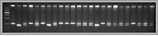 1. Objectives The GMCC will provide essential resources that will enable the research community to greatly improve the speed and efficiency of mapping genes that modulate the size and cell composition of the mouse brain. We will generate close to 400,000 genotypes for a set of approximately 1400 mice (1200 animals between the ages of 46 and 76 days, and ~200 animals more than 2 years old) during this grant period. These genotypes will be a vital component of the Neurogenetics Tool Box. The brains of mice that have been genotyped by this core will be processed by the Neurohistology Core, and high-resolution images of all the cases will be added to the Mouse Brain Library (MBL). Genotype databases will be consolidated with the quantitative data generated as part of the MBL and the NeuroCartographer Project (Project 3). When this work is complete, the Neurogenetics Tool Box will contain a huge body of phenotypic and genotypic data for a population of over 1400 adult animals from a unique type of genetic cross. These data will make it possible to routinely map QTLs with a precision of <4 cM. The GMCC will also be responsible for oversight of our mouse colony and for providing fixed brains to the Neurohistology Core. The maintenance of the mouse colony is not otherwise discussed in this Core description because these duties will be covered primarily by the UT Memphis Department of Comparative Medicine. Intramural funds provided by the Department of Anatomy and Neurobiology will cover minor expenses associated with perfusion and dissection of brains. We routinely process several thousand cases per year as part of other funded projects that deal with the structure of the mouse eye and retina. Additional supplies or technical assistance will not be required for the duration of this grant period. Current
status. A
high-resolution mapping panel. The tenth-generation BD intercross we are using has many advantages over a conventional intercross or backcross. With a conventional intercross or backcross, QTLs can generally be mapped only with a precision of 10 cM, even when 2000 or more cases are studied. To overcome this problem, Darvasi (1998) suggested the advanced intercross as a conceptually simple method to increase the precision of QTL mapping to 2 cM. An advanced intercross accumulates recombination events over many generations (see below), effectively stretching the genetic map. The genetic map doubles in length with each doubling of generation number. For example, a G4 cross has twice the map length of an F2 cross. The genetic map of G10 animals is five times as long as the map based on a conventional cross7000 cM rather than 1400 cM. This means that for a given level of effort, the G10 panel provides five times the precision of a F2 intercross. Thus, positional candidate gene cloning approaches become far more feasible. Dr. K. Manly has recoded the Map Manger QT program to create Map Manager QTX, which will accommodate data sets from advanced intercrosses (Manly and Olson 1999). Project 4, the Neurogenetics Tool Box, will incorporate both genotypes and quantitative neuroanatomical traits from the entire G10 panel. Advantages
for Neuroinformatics. Statistical
Power. 2.
Staffing and Oversight
The GMCC will be staffed by one full-time professional. Dr. Jing Gu will be responsible for the extensive genotyping. Dr. Gu currently runs the PIs genotyping lab. Dr. Gu will also be responsible for error checking and for entry of genotype data into our consolidated relational databases. She will be supported by the PPG and will be responsible for genotyping the G10 advanced intercross. She is highly skilled in all aspects of genotyping. The Molecular Resources Center will provide additional personnel to assist with the work. 3.
Resources and Environment
The Genotyping and Mouse Colony Core will be housed at the University of Tennessee, Memphis. This Core will initially share facilities in the PIs lab (311 Wittenborg). However, late in Year 01 we will open a new UT Memphis Genotyping Center as a division of the UT Memphis Molecular Resources Center. The Genotyping Center will be directed by Dr. Robert Williams. This facility will have an initial capacity of at least 200,000 genotypes a year, of which half will be devoted to this PPG (see letter from Dr. Michael Dockter, Vice Dean for Research). 4.
Administration
Dr. Jing Gu will report to Drs. Williams and Manly. Drs. Williams and Gu will meet at least once a week to review progress and will keep closely in touch with Dr. Manly by email and phone. Dr. Gu will maintain several key databases that will be used by Dr. Manlys group as part of Project 4. Dr. Manly will have direct access to the FileMaker Pro server that will host the genotype databases. Funds for the Genotyping and Mouse Colony Core will be managed by the Department of Anatomy and Neurobiology and by the Administrative Core of the Program Project. Funds to buy equipment to support the expanded genotyping throughput will be provided by the University of Tennessee. The limited support required to fix animals and ship brains to Dr. Rosen will be covered by intramural funds. The budget for this core does not include any equipment request. Our supplies request is limited to genotyping (~$0.20/genotype) and is relatively modest. Our cost is far lower than current commercial genotyping prices. For example, Research Genetics currently charges in excess of $4 per microsatellite genotype. We will be able to generate 400,000 genotypes for well under $80,000 in supplies. 5.
Justification Full-scale genotyping and extension of the Mouse Brain Library, as proposed in this application, will allow multiple research groups to study many traits in the same set of animals, maximizing the utility of each cross, preventing duplication of effort, and revealing the complex genetic basis of variation in different CNS compartments. This community effort also allows us to substantially increase the positional precision of QTLs that are mapped. The proposed project will greatly reduce the cost and effort of examining the basis of normal variation in CNS architecture among and within strains of mice. Until now, it has been possible to adequately analyze only a small number of phenotypes in each animal that is genotyped. Each research group generated a unique F2 intercross or backcross consisting of several hundred animals. Researchers observed a small number of traits, genotyped their modest sample of animals, and subsequently mapped the loci modulating the observed traits. R. Williams and colleagues have used this approach to map more than a dozen QTLs that control morphometric and quantitative variation in the architecture of the eye and brain of normal strains of mice (see Appendix; Williams et al. 1998; Zhou and Williams 1999; Williams 2000). Only four trait values were acquired for most mice: body weight, brain weight, eye weight, and retinal ganglion cell number. Even this was a huge undertaking for a single lab. This cottage industry approach is incredibly inefficient and yields only low-resolution estimates of the chromosomal location of QTLs and their phenotypic effects. Use
of the core by individual projects. 6.
Procedures
Generating
the AI cross. Scanning
the genome for QTLs. Genotyping
the AI cross. Data
entry. Fixation. 7.
Financial Considerations
Technical
support.
A full-time senior technician is needed to carry out activities
that are part of this Core. Equipment.
The Vice Dean of Research at UT Memphis will provide $140,000 for
equipment required to perform as many as 400,000 genotypes over the
duration of this grant. The PI will use these funds to assemble a new
high-throughput genotyping lab. The appended letter from the Dean lists
some of the key equipment that will be purchased. Travel costs. None 8.
Bibliography (Core B) Darvasi
A, Soller M (1995) Advanced intercross lines, an experimental population
for fine genetic mapping. Genetics 141:11991207. Darvasi
A (1998) Experimental strategies for the genetic dissection of complex
traits in animal models. Nat Genet 18:1924. Dietrich WF, Miller JC, Steen RG,
et al. (1994) A genetic map of the 4006 simple sequence length
polymorphisms. Nat Genet 7:220245Doerge RW,
Churchill GA (1996) Permutation tests for multiple loci affecting a
quantitative character. Genetics 142:285291. Manly,
K.F. and Olson, J.M. (1999)
Overview of QTL mapping software and introduction to Map Manager QT. Mamm
Gen 10: 327-334. Roff DA
(1997) Evolutionary quantitative genetics. Chapman Hall, New York. Schmitz
S., Cherny SS, Fulker DW (1998) Increase in power through multivariate
analyses. Behav Genet 28:357363. Williams
RW (1998) Neuroscience meets quantitative genetics: Using morphometric
data to map genes that modulate CNS architecture. In: Morrison J, Hof P (eds)
Short course in quantitative neuroanatomy. Society of Neuroscience,
Washington DC, pp 6678. Williams
RW (2000) Mapping genes that modulate mouse brain development: A
quantitative genetic approach. In: Goffinet A, Rakic P (eds) Mouse Brain
Development. Springer, Berlin (2000) in press. Zhou G,
Williams RW (1999) Eye1 and Eye2:
Gene loci that modulate eye size, lens weight, and retinal area in mouse.
Invest Ophthalmol Vis Sci 40:817825. e.
Human Subjects
none f.
Vertebrate
Animals Description
of proposed use of animals i. We will use the species Mus domesticus in these studies. Most animals are originally obtained from the Jackson Laboratory, Bar Harbor, Maine. Both sexes will be used. We will have breeding colonies and intercrosses of selected strains. We will have a colony of aged mice to study the genetics of CNS aging. ii. Justification of animal use. Mice will be used to characterize the genetics of CNS. Over the period of this grant, we anticipate that we will use an average of approximately 1000 mice per year. Such large numbers of animals are required in order to map quantitative trait loci with precision. All of these animals will be generated in our mouse colony. iii. Veterinary care. The mice will be maintained in departmental AAALAC approved facilities under the supervision of the Department of Comparative Medicine of the University of Tennessee. Only trained personnel will work with our mouse colony, and they will check room conditions, cage, and animal status daily. The Department of Comparative Medicine has new facilities in the Nash Annex. This is a state-of-the-art vivarium with extensive space for pathogen-free colonies of mice. All of our colony will be kept in this facility. The facility employs two full-time veterinarians to monitor the health and well-being of animals. iv. Analgesics and anesthetics. NA v. Euthanasia method. Adult mice that will be fixed by transcardial perfusion will be anesthetized deeply with Avertin. Adult mice that will be used without perfusion will be sacrificed by cervical dislocation. Neonates and fetal animals are decapitated with scissors. Some mice will be euthanized by exposure to 100% CO2. These methods are consistent with the recommendations of the Panel on Euthanasia of the American Veterinary Medical Association. i.
Consultants Dr. Warren Young (Scripps Institute) will serve as a consultant on
Projects 1 . |
|||
|
REFERENCES |
|||
|
|||