 |
| |||||
|
| ||||||||
|
| ||||||||
Home  Publications Publications |
|
|
Growth Cones and Dying Axons This monster took the better part of three years to complete. The web edition has been updated with new figures (including color-tinted electron micrographs). If you would like to see high-resolution images of growth cones and dying axons, this is a great source. The paper was published in The Journal of Comparative Neurology in 1986 by R. W. Williams, Michael Bastiani, Barry Lia, and Leo Chalupa. We show that about 80% of all retinal ganglion cells die early in development in domestic cats. The paper has a fairly long discussion on the developmental significance (and insignificance) of neuron death. 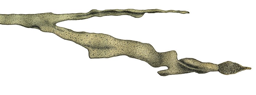 The terminal 18 µm of a typical growth cone (ID #22) from the E39 monkey optic nerve. The total volume of the large branch is .9 µm3, and its surface area is 54 µm2. At its tip is an aggregate of vesicles that we believe are associated with a transient retraction at or during during fixation. The distant branch is in a different fascicle and has one-half the surface area of the major branch. (Reconstruction shown from sections 300 to 501.) Do growth cones like the one illustrated above track along pre-existing axons as they grow backward into the thalamus and midbrain? The short answer, described in this 1985 paper in Proceedings of the National Academy of Science paper, is No they don't. I spent eight months photographing, tracing, and retracing a set of several hundred embyronic axons through 500 serial electron micrographs. IThe first time I ever gave a research seminar I described this grueling work and its significance. At the end of my talk a faculty member in the audience let me know that this work was of no signficiance whatsover because I had only studied a single E39 embryo. Over the next few years I have paid penance for my "N of 1" sin. It is still a fun paper to look at. In this web edition I have added numerous color figures and overlays that illustrate the meandering paths taken by growth cones. A very detailed quantitative analysis of growth cone traffic patterns in the optic nerve. Growth cones do not express a strong preference for glial cells or the basal lamina. This paper with Pasko Rakic was published in The Journal of Neuroscience in 1991. We have added several new color figures to this web edition, and new data on growth cone distribution in the optic chiasm. |
|

Neurogenetics at University of Tennessee Health Science Center
| Top of Page |
Mouse Brain Library | Related Sites | Complextrait.org
